News from Augencenter Wollishofen (Zurich)
Learn more about news from the Augencenter Wollishofen and answers to many patient questions.
News from Augencenter Wollishofen

Should I go to the eye doctor even if I have no problems with my vision?
From the age of 40, annual examination, especially of the optic nerve and eye pressure, is recommended. In this way, glaucoma can be ruled out. It is important to detect eye diseases early and to ...
Does my child need glasses?
In children, it is extremely important to detect vision problems early and treat them correctly. The brain learns to see in the first years of life. Problems that are not treated by the time a child starts school ...
Why are my eyes watering?
The causes are diverse and often multifactorial. Foreign bodies in the eye or small injuries of the cornea, as well as inflammations and allergies lead to increasing tear production. On the one hand, foreign bodies are thus flushed out and on the other ...
Does it harm the eye to read in poor light?
For adults it is not harmful but exhausting and tiring. For children, a possible damage is discussed. However, there is no clear scientific evidence.

If you have eye flashes, go to the ophthalmologist?
In children, it is extremely important to detect vision problems early and treat them correctly. The brain learns to see in the first years of life. Problems that are not treated by the time a child starts school ...
Slow down presbyopia with glasses?
No. Presbyopia is caused by the normal aging process of the lens. It becomes more and more rigid and thus the ability to see well at close range is lost. This process is not ...

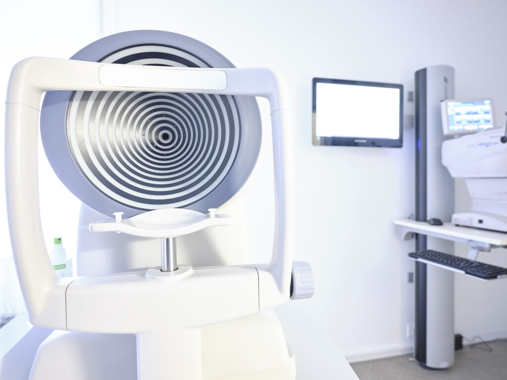
Examine visual field
Visual field determination is used primarily for early detection of glaucoma. During the visual field examination (perimetry), the examination of the outer and inner limits of the visual field as well as the sensitivity of the visual function outside the ...
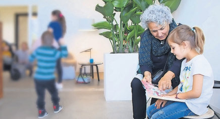
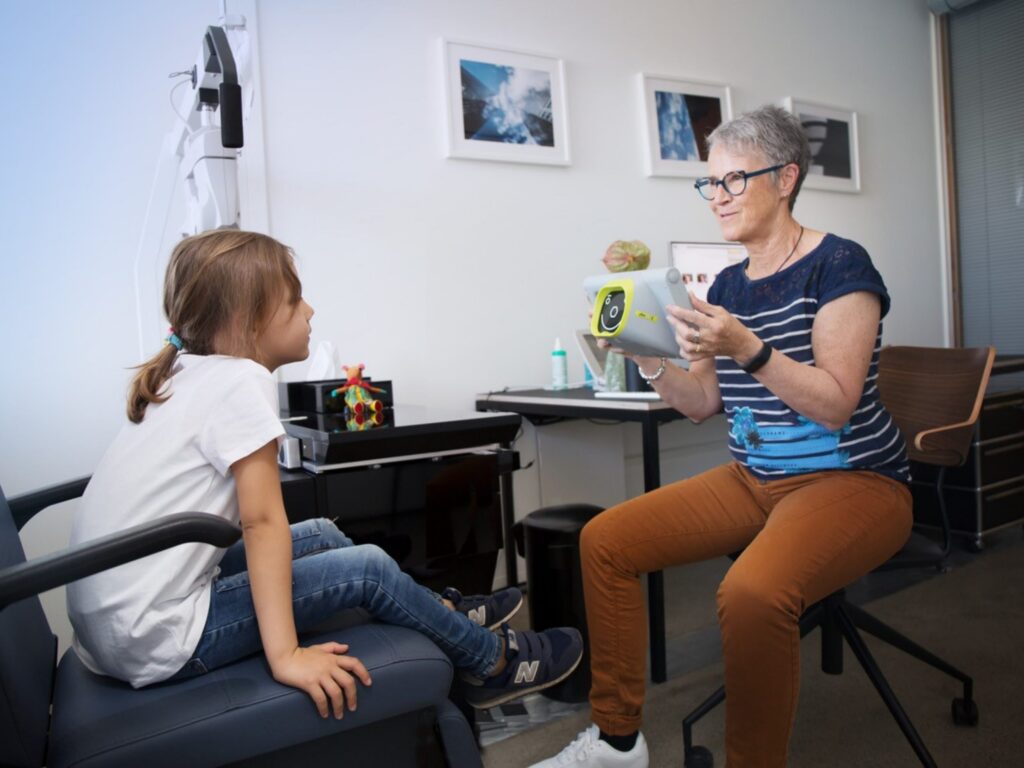
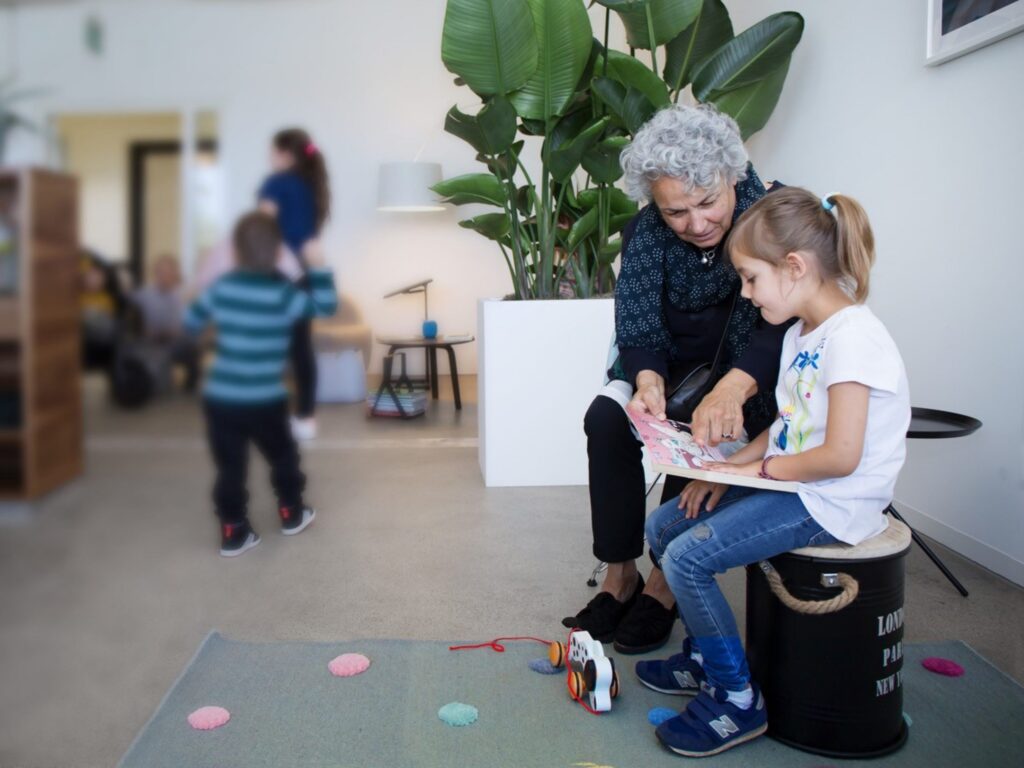
Augencenter Wollishofen: Where children like to go to the eye doctor !!!
A modern ophthalmology practice in Wollishofen is dedicated to the well-being of its young patients. The children are involved in the examination and thus motivated to participate. On this Thursday afternoon, the atmosphere in the ...

Why good vision is so important
If one sense fails, the other senses must additionally take over its work. The eyes play a special role in this. The human senses are the contact to the environment. The human ...
Examine optic nerve
A healthy optic nerve head curves slightly inward. The optic nerve transmits our sensory impressions from the eye to the brain. Vessels run through its center to supply the retina. The optic disc, the ...
Examine intraocular pressure
During the screening of glaucoma, intraocular pressure measurement (tonometry) is used and the optic disc is assessed. The examination can determine whether the intraocular pressure has increased and suspected ...
Examine anterior eye segments
In ophthalmology, the eye is divided into two sections: The anterior and posterior sections of the eye. This section includes the optics of the eye: conjunctiva, cornea (cornea), iris (iris)...
Ophthalmoscopy: Examine the fundus of the eye (funduscopy)
Examination of the fundus of the eye is a most important diagnostic procedure. With an ophthalmoscope it is possible to look through the pupil to the back of the eye and see the retina , the optic nerve, the ...
Blind spot (papilla)
The so-called blind spot is the point in the eye where the retina is interrupted and merges with the optic nerve. Since there are no light receptors at this point, ...
Inner eye shell
The inner shell forms the retina. It is used to receive light stimuli. The light impressions are transmitted to the brain via the optic nerve and processed there.
Optic nerve (lat. Nervus opticus) in the eye
The optic nerve is responsible for ensuring that information from the retina reaches the brain. The concentrated bundle of millions of nerve fibers converts the light stimuli from the retina into impulses and sends them to ...
Yellow spot (lat. Macula lutea) in the eye
The yellow spot is located in the center of the retina and is only about three to five millimeters in size. It gets its name from a yellow pigment that is found on this spot. ...
Retina (lat. Retina)
The retina is responsible for converting light entering the eye into nerve impluses. The retina is very sensitive to light and lines the inside of the eye. It is the largest retina in the world with approximately 127 ...
Vitreous body (lat. Corpus vitreum)
The vitreous body fills most of the interior of the eye and thus sits between the lens and the retina. Consequently, light entering through the pupil and lens must pass through the vitreous before ...
Blood vessels in the eye
While the retina is supplied by the choroid from one side, blood vessels also lie directly on the back of the eye - the retina. This network of vessels comes ...
Choroid (lat. Choroidea)
The choroid is located between the sclera and cornea and extends over the entire posterior part of the eyeball. It has a high density of blood vessels and supplies the retina with ...
Sclera (lat. Sclera) in the eye
The sclera forms the outermost layer of the eye, it is "the white of the eye" that can be seen from the outside around the pupil and iris. It is therefore also called ...
External eye muscles
The external eye muscles are responsible for the movements of the eyes, i.e. changing the direction of gaze. They attach to different parts of the eyeball. Humans have four straight and two ...
Lenticular bands (lat. Zonula ciliaris)
The lens ligaments - also called zonular fibers - are elastic fibers arranged in a spoke-like pattern around the capsule in which the lens of the eye is located. They hold the lens in position ...
Eye lens (lat. Lens crystallina)
The lens of the eye focuses the light entering through the pupil. With approximately 10 to 20 diopters of the eye's total refractive power (of about 63 diopters), the lens of the eye - along with the cornea ...
Middle eye shell
The middle shell consists of three sections with specific functions. The iris adjusts the incidence of light. The ciliary body produces aqueous humor and regulates the curvature of the lens for near or ...
Rainbow skin (lat. Iris)
The iris is the colorful part of the eye and is as individual as a fingerprint. The so-called iris, or "colorful aperture" of the eye, can be changed by an individual pigment distribution in ...
Anterior chamber of the eye (lat. Camera anterior bulbi)
The anterior chamber is larger than the posterior chamber and is the area of the eye from the back of the cornea to the iris. Through a small gap between ...
Pupil (lat. pupilla): Eye hole of the eye
The pupil is the circular aperture in the center of the iris. Light enters the interior of the eye through the pupil. Its size is regulated by the surrounding iris, depending ...
Cornea (lat. Cornea)
Light enters the eye through the cornea, where it is then transmitted to the lens and pupil. The cornea is therefore also called the "window of the eye" and is a ...